
Scientifically reviewed by
Dr. Ky H. Le, MD
Disclaimer: The information presented in this article is intended for educational and research purposes only, aimed at laboratory professionals, researchers, and collaborators. This content does not constitute medical or clinical advice.
Cerluten is a unique complex of peptide bioregulators extracted from cerebral cortex tissue. Unlike synthetic peptides, which typically act through receptor-mediated signaling, these brain-specific compounds interact directly with cellular DNA, regulating gene expression and protein synthesis.
The effects of Cerluten have been studied in laboratory settings and clinical trials. Laboratory studies have shown relevant data on neuronal differentiation, antioxidant properties, and the prevention of synaptic loss. Clinical trials have provided objective measurements of brain function, offering a comprehensive view of Cerluten’s potential benefits for cognitive health.
What Is the Cerluten Peptide Bioregulator?
Cerluten is a peptide bioregulator extracted from the brain tissue of young animals, specifically from the cerebral cortex of individuals under 12 months of age. The extraction process yields low molecular weight peptides (up to 10,000 Da) that naturally regulate neuronal metabolism and function.
This peptide complex exhibits specific activity concentrated in neurons and glial cells. Compounds derived from Cerluten are capable of crossing the blood-brain barrier when administered peripherally, enabling targeted effects in the central nervous system (CNS) without broad systemic activity.
Differences Between Bioregulators and Standard Peptides
The defining characteristic of bioregulators lies in their mechanism of action. Standard synthetic peptides typically bind to receptors on the cell surface to trigger signaling cascades.
In contrast, bioregulators penetrate cellular and nuclear membranes, directly interacting with DNA and chromatin structures. This interaction allows them to modulate transcriptional activity through complementary binding to gene promoter regions.
Brain-derived bioregulators demonstrate high selectivity for binding sites within genes associated with neuronal function. Molecular modeling studies suggest preferential interaction with specific nucleotide sequences.
Additionally, these peptides present significant therapeutic potential, particularly in neuroprotection and regenerative contexts.
Molecular Mechanisms of Action
Gene Regulation via DNA Interaction
Cerluten peptides bind to specific DNA sequences located in gene promoter regions through sequence-specific recognition. These short peptides show particular affinity for regions containing charged nucleotides.
Their mechanism involves interactions with both double-stranded DNA and histone proteins (H1, H2B, H3, H4). This binding alters chromatin accessibility, promoting increased transcription of target genes.
Epigenetic modulation occurs at concentrations ranging from 2 to 200 ng/mL, demonstrating high potency compared to traditional signaling peptides.
Protein Synthesis and Cellular Metabolism
Brain peptide bioregulators regulate the expression of genes encoding proteins essential for neuronal structure and function. Research has documented increased expression of neurogenesis markers, including Nestin, GAP43, β-tubulin III, and doublecortin, in response to these short peptides.
These effects translate into enhanced protein synthesis supporting neuronal differentiation, synaptic plasticity, and cellular repair processes. Additionally, peptides influence the expression of neurotrophic factors such as NGF and BDNF, along with their respective receptors.
Metabolic regulation occurs through modulation of genes directly linked to mitochondrial function, including components of the electron transport chain and ATP synthesis mechanisms.
Tissue-Specific Activity
The preferential activity observed in central nervous system (CNS) cell populations reflects the origin of these peptides from cerebral cortex tissue. Brain-derived peptides exhibit regulatory effects focused on neurons and glial cells, minimizing systemic effects.
This tissue specificity enables targeted modulation of neuronal gene expression patterns, regulating metabolic processes characteristic of brain tissue without interfering with other organ systems.
Oxidative Stress and Antioxidant Response
Brain peptide bioregulators modulate the expression of antioxidant enzymes through multiple signaling pathways. Studies indicate increased levels of superoxide dismutase (SOD2), glutathione peroxidase (GPX1), and catalase.
These peptides interfere with oxidative mechanisms in the cerebral cortex, reducing lipid peroxidation and protein oxidation. Research reports decreased levels of oxidative stress biomarkers such as malondialdehyde and 4-hydroxy-2-nonenal in treated tissues.
Additionally, these compounds regulate genes responsible for mitochondrial quality control, reducing oxidative stress at the primary site of reactive oxygen species production.
Neurogenesis and Neuronal Differentiation
Short peptides similar to those found in Cerluten stimulate neuronal differentiation from stem cells. These bioregulators activate genes essential for neuronal development, including transcription factors and structural proteins.
Experiments show that peptides such as KE, AED, KED, and AEDG increase the expression of GAP43, a marker of synaptic plasticity and axonal growth. Treatment also enhances Nestin expression, indicating activation of neural stem cells.
Peptides promote dendritic branching, increasing both the number of primary dendrites and total dendritic length in cultured neurons, supporting improved neuronal connectivity.
Synaptic Function and Plasticity
Neuroprotective peptides maintain dendritic spine density in neurodegeneration models. Sequences such as EDR and KED prevent the loss of these structures in neuronal cultures exposed to β-amyloid toxicity.
These agents regulate genes encoding proteins essential for synaptic architecture and function, including components of the postsynaptic density and adhesion molecules.
By preserving dendritic spine morphology, bioregulators support synaptic plasticity mechanisms crucial for learning and memory consolidation.
Prevention of Apoptosis
Brain peptides attenuate apoptotic pathways through multiple mechanisms. They suppress pro-apoptotic factors such as caspase-3 and p53 while preserving anti-apoptotic proteins.
In models of cerebral ischemia and neurodegenerative diseases, peptide analogs normalize the activation of stress-induced kinases (SAPK/JNK, pERK1/2), which are involved in neuronal death cascades.
This anti-apoptotic action supports neuronal survival under conditions of metabolic stress, excitotoxicity, and oxidative damage.
Neurophysiological Changes
Electroencephalographic (EEG) analysis has demonstrated significant improvements in brain bioelectrical activity. Participants showed transitions from pathological EEG patterns (types III, IV, and V) to normalized configurations.
Type III patterns, characterized by low-amplitude and irregular activity, improved in 7 out of 11 individuals. Type IV patterns, marked by excessive rhythm regularity, normalized in 6 out of 10 cases. Type V patterns, with irregular slow waves and elevated paroxysmal spikes, improved in 7 out of 15 patients.
The alpha index, which measures the intensity of the dominant rhythm, showed the following values:
| Group | Pre-treatment | Post-treatment |
| Cerluten | 34.0 ± 4.1 | 47.9 ± 3.7 |
| Control | 33.6 ± 3.7 | 41.3 ± 4.2 |
These data indicate a 41% improvement in the peptide-treated group compared to 23% in the control group. The peptides enhanced alpha rhythm modulation, restored zonal EEG variations, and reduced irritative processes.
Nota: Para enriquecer visualmente nosso conteúdo, informamos que algumas das imagens utilizadas em nossos artigos são criadas por meio de inteligência artificial. Elas servem a propósitos puramente ilustrativos e não devem ser interpretadas como representações de eventos ou pessoas reais.
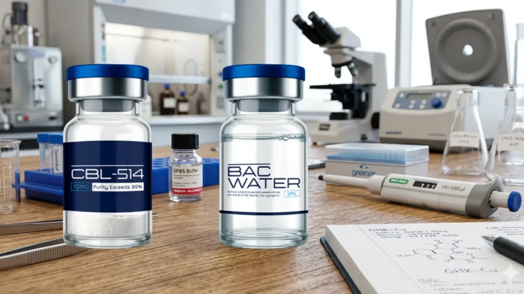
TECHNICAL AND SCIENTIFIC GUIDE CBL-514: Pharmacology, Clinical Evidence and Therapeutic Outlook Updated 29 July 2026 Intended audience: researchers,...
Read more...
1. Abstract Lyophilization, or freeze-drying, is the most effective method for stabilizing both synthetic and natural peptides. By...
Read more...
1. Abstract Ovágeno is a synthetic tripeptide (L-glutamic acid, L-aspartic acid, and L-proline) developed to act as a...
Read more...
1. Nature and Molecular Architecture AOD9604 is a synthetic peptide analog that mimics the C-terminal region of human...
Read more...
BIOPELABS delivers research-focused peptides with a clean, modern experience—clear product options, responsive support, and secure shopping. Explore our peptide catalog and find the right format for your needs today.
Disclaimer: The products offered by BiopeLabs are intended for research and laboratory use only. They are not approved for human consumption, therapeutic, diagnostic, or veterinary use. By purchasing from our website, you acknowledge that you are a qualified researcher or purchasing on behalf of a legitimate research institution and agree to use these products in accordance with all applicable laws and regulations.

1: 5-Amino-1MQ 10mg
2: 5-Amino-1MQ 50mg
3: ACE-031 1mg
4: ACTH 1-39 5mg
5: Adamax 5mg
6: Adamax 10mg
7: AHK-Cu 50mg
8: AICAR 50mg
9: AICAR 100mg
10: AOD-9604 5mg
11: AOD-9604 10mg
12: Alprostadil 20mcg
13: Amylin 5mg
14: Angiotensin (1–7) 5mg
15: ARA-290 10mg
16: Bimagrumab 5mg
17: Botulinum Toxin 100IU
18: BPC-157 10mg
19: BPC-157 20mg
20: BPC-157 5mg + TB-500 5mg
21: BPC-157 10mg + TB-500 10mg
22: Bronchogen 20mg
23: Cagrilintide 10mg
24: Cagrilintide 20mg
25: Cardiogen 20mg
26: Cartalax 20mg
27: CBL-514 10mg
28: CBL-514 20mg
29: CBL-514 30mg
30: Cerebrolysin 60mg
31: Cerlankin 2mg
32: CGRP (Calcitonin Gene-Related Peptide) 1mg
33: CJC-1295 with DAC 5mg
34: CJC-1295 with DAC 10mg
35: CJC-1295 with DAC 5mg + Ipamorelin 5mg
36: CJC-1295 without DAC 5mg
37: CJC-1295 without DAC 10mg
38: CJC-1295 without DAC 5mg + Ipamorelin 5mg
39: Chonluten 20mg
40: Cortagen 20mg
41: Crystagen 20mg
42: CT-1 Cardiotrophin-1 5mg
43: Dermorphin 5mg
44: Dihexa 5mg
45: DSIP 5mg
46: Dulaglutide 5mg
47: Elabela Apelin 5mg
48: Enfuvirtide 90mg
49: Epithalon 5mg
50: Epithalon 40mg
51: Epithalon 50mg
52: EPO 3000IU
53: FGF21 10mg
54: Follistatin-344 1mg
55: FOXO4 10mg
56: GHK-Cu 50mg
57: GHK-Cu 100mg
58: Ghrelin 10mg
59: GHRP-2 5mg
60: GHRP-6 5mg
61: Glucagon 1mg
62: Glutathione 1500mg
63: Gonadorelin Acetate 5mg
64: Goserelin 10mg
65: GLOW BPC-157 10mg + GHK-Cu 50mg + TB-500 10mg
66: HGH 191AA 15IU
67: HGH 191AA 36IU
68: HGH Fragment 176-191 10mg
69: HGH Fragment 176-191 15mg
70: HMG (Human Menopausal Gonadotropin) 75IU
71: Humanin 10mg
72: Hyaluronic Acid 5mg
73: Hexarelin Acetate 2mg
74: Hexarelin Acetate 5mg
75: IGF-1 LR3 1mg
76: Ipamorelin 5mg
77: Ipamorelin 10mg
78: Kisspeptin-10 5mg
79: Kisspeptin-10 10mg
80: KLOW BPC-157 10mg + GHK-Cu 50mg + TB-500 10mg + KPV 10mg
81: KPV 10mg
82: Leuprolide Acetate 10mg
83: Liraglutide 5mg
84: Livagen 20mg
85: LL-37 5mg
86: Lysyl Oxidase 30mg
87: Matrixyl 10mg
88: Mazdutide 10mg
89: Melanotan I 10mg
90: Melanotan II 10mg
91: Melatonin 10mg
92: MGF 2mg
93: MOTS-c 10mg
94: MOTS-c 40mg
95: NAD+ 100mg
96: NAD+ 500mg
97: NAD+ 1000mg
98: Nesiritide 5mg
99: Neuropeptide Y (NPY) 10mg
100: Ovagen 20mg
101: Oxytocin Acetate 5mg
102: Oxytocin Acetate 10mg
103: P21 5mg
104: P21 10mg
105: PACAP 5mg
106: Pancragen 20mg
107: PE 22-28 10mg
108: PEG-MGF 2mg
109: Pinealon 5mg
110: Pinealon 10mg
111: Pinealon 20mg
112: PNC-27 5mg
113: PNC-27 10mg
114: Prostamax 20mg
115: PT-141 10mg
116: PTH Fragments 10mg
117: Relamorelin 5mg
118: Retatrutide 10mg
119: Retatrutide 20mg
120: Retatrutide 30mg
121: Retatrutide 40mg
122: Retatrutide 60mg
123: Retatrutide 5mg + Cagrilintide 5mg
124: Selank 5mg
125: Selank 10mg
126: Semaglutide 5mg + Cagrilintide 5mg
127: Semax 5mg
128: Semax 10mg
129: Sermorelin 5mg
130: Sermorelin 10mg
131: SLU-PP-332 10mg
132: SNAP-8 10mg
133: SS-31 10mg
134: SS-31 50mg
135: Survodutide 10mg
136: TB-500 5mg
137: TB-500 10mg
138: TB-500 (FRAG) 10mg
139: Teduglutide 5mg
140: Teriparatide 10mg
141: Tesamorelin 5mg
142: Tesamorelin 10mg
143: Tesamorelin 20mg
144: Tesamorelin 5mg
145: Tesofensine 1mg
146: Testagen 20mg
147: Thymalin 10mg
148: Thymosin Alpha-1 5mg
149: Thymosin Alpha-1 10mg
150: Treprostinil 5mg
151: Vesugen 20mg
152: VIP 5mg
153: VIP 10mg
154: Vilon 20mg