
Scientifically reviewed by
Dr. Ky H. Le, MD
Disclaimer: The information presented in this article is for educational and research purposes only, intended for laboratory professionals, researchers, and collaborators. This content does not constitute medical or clinical advice.
KPV (Lys-Pro-Val) is a low molecular weight tripeptide sequence derived from the melanocyte-stimulating hormone (α-MSH), recognized for its significance in research related to inflammation and epithelial barrier function. This C-terminal fragment retains several bioactive properties of the original hormone, while its smaller size and enhanced cellular penetration capabilities make it a practical choice for laboratory investigations.
Scientists working in fields such as inflammatory signaling, epithelial transport, and host defense often choose KPV as a compact molecular probe. Its interactions with NF-κB signaling pathways, efficient passage through cellular membranes with the aid of specific transporters, and involvement in nuclear import processes support its use across a wide range of in vitro experiments.
KPV is a tripeptide sequence formed by amino acid residues 11 to 13 of α-MSH, constituting the C-terminal fragment of this hormone. While α-MSH primarily activates melanocortin receptors due to its central sequence HFRW, KPV follows a distinct pathway.
Instead of relying on these receptors, KPV operates through mechanisms that do not involve receptors. Surprisingly, these pathways still reproduce many of the anti-inflammatory effects associated with α-MSH.
When comparing full-length α-MSH with its shorter fragments, researchers find that KPV represents the shortest sequence necessary to preserve important biological properties. If a peptide does not contain this specific sequence, it quickly loses much of its anti-inflammatory and antimicrobial activity in experimental models.
By analyzing the sequence, composed of lysine, proline, and valine, a combination of charged and hydrophobic characteristics is noted. Due to this amphipathic nature, KPV can interact with cellular membranes and is rapidly recognized by transporters.
Classical melanocortin signaling elevates cAMP through G-protein-coupled receptors. However, KPV diverts from this model, producing biological effects in cells where cAMP pathways are pharmacologically inhibited.
Calcium signaling serves as an alternative pathway. In keratinocyte models, KPV induces rapid increases in intracellular Ca²⁺, independent of cAMP, suggesting multiple modes of signaling for this peptide.
KPV is small enough to penetrate cells using transport systems inaccessible to larger proteins. This helps explain KPV’s biological activity.
The proton-coupled oligopeptide transporter, PepT1 (SLC15A1), serves as the primary entry route for KPV into the intestinal epithelium. This transporter typically carries dietary di- and tripeptides from the intestinal lumen into epithelial cells.
Studies employing PepT1 inhibitors or genetic knockdown demonstrate a reduction in KPV uptake and subsequent decreased effects on inflammatory signaling. Competitive substrates for PepT1 also inhibit intracellular accumulation of KPV.
PepT1 expression increases during inflammation in the colonic epithelium, potentially enhancing KPV transport precisely when anti-inflammatory signaling is most needed to maintain the barrier.
Time-lapse imaging of human bronchial epithelial cells reveals that KPV transitions from cytoplasmic to nuclear localization within hours of absorption. Immunofluorescence studies using histidine-tagged KPV show fluorescence concentrated in the nuclei.
This nuclear accumulation occurs alongside the suppression of NF-κB activation, suggesting the presence of direct nuclear mechanisms, rather than merely cytoplasmic effects.
Nuclear import of NF-κB subunits requires importin proteins that recognize nuclear signaling sequences. Competition assays indicate that KPV interferes with the binding of p65RelA to importin-α3, specifically in the domain that recognizes the nuclear localization signal of p65.
This molecular interaction explains how the nuclear presence of KPV correlates with its effects on NF-κB signaling. By occupying importin binding sites, the peptide prevents the nuclear entry of p65.
NF-κB is central to controlling the production of inflammatory proteins by cells. KPV can impact this pathway at various stages, elucidating many of its observed effects in epithelial cell studies.
In unstimulated cells, IκBα sequesters NF-κB dimers in the cytoplasm. Inflammatory stimuli induce phosphorylation and degradation of IκBα, releasing NF-κB for nuclear translocation.
Treatment with KPV elevates total IκBα levels after TNF-α stimulation without altering the ratio of phosphorylated to total IκBα. This pattern suggests reduced degradation of IκBα, preserving cytosolic sequestration of NF-κB through a post-phosphorylation mechanism.
Live-cell imaging with fluorescently tagged p65RelA shows KPV’s ability to block nuclear import. KPV-pretreated cells maintain low p65 ratios between nucleus and cytoplasm, even after TNF-α exposure, while control cells show rapid nuclear accumulation.
This blockage occurs at the importin level, corroborating competition data. Nuclear levels of p65 determine transcriptional activity, making it a critical control point.
Several studies confirm reduced NF-κB transcriptional activity following KPV treatment:
These results reflect the pathway’s role in the production of inflammatory mediators across various tissue types.
KPV’s effects may vary between different epithelial lineages, reflecting differences in transporter expression, inflammatory signaling architecture, and baseline NF-κB activity.
Intestinal research systems focus on PepT1-dependent mechanisms. Experiments with Caco-2 cells and primary intestinal epithelium show that PepT1 expression levels correlate with KPV’s anti-inflammatory potency.
Barrier function studies report preservation of tight junction integrity under inflammatory stress. Changes in transepithelial electrical resistance and permeability markers indicate maintenance of barrier competence.
Coculture models including immune cells show reduced secretion of pro-inflammatory cytokines in both epithelial and immune layers, suggesting that KPV has effects on multiple cell populations.
Research with human bronchial epithelial cells provides important mechanistic insights. Studies in these systems characterize:
These findings position KPV as a protective agent for epithelial proliferation and matrix remodeling control.
Skin models reveal that calcium signaling pathways are distinct from cAMP-dependent mechanisms. KPV induces rapid Ca²⁺ increases in keratinocytes, even when adenosine receptor agonists block cAMP elevation.
Assays using NF-κB reporters and DNA binding studies confirm transcriptional suppression similar to that observed in other epithelial types. The combination of calcium signaling and NF-κB modulation suggests multiple competitive pathways in the skin.
In addition to its anti-inflammatory properties, KPV also exhibits antimicrobial activities, placing it in the category of host defense peptides.
α-MSH demonstrates activity against Candida albicans, Cryptococcus neoformans, and Staphylococcus aureus, including methicillin-resistant strains. Fragment studies identify the KPV sequence as essential for anti-staphylococcal effects. Peptides lacking this sequence lose antimicrobial activity.
Expanded fragments including KPV show direct antimicrobial and antifungal actions in plate assays, with effects appearing to be associated with membrane disturbances and changes in microbial intracellular signaling.
Chemical modifications are employed to investigate sequence requirements for antimicrobial function. Glycoalkylation or acetylation of the lysine residue eliminated antimicrobial activity under some testing conditions, highlighting the importance of native charge distribution.
These data on structure and activity indicate membrane-sensitive or interface mechanisms, typical of antimicrobial peptides. Minor chemical alterations can affect the amphipathic balance necessary for microbicidal effects.
Nota: Para enriquecer visualmente nosso conteúdo, informamos que algumas das imagens utilizadas em nossos artigos são criadas por meio de inteligência artificial. Elas servem a propósitos puramente ilustrativos e não devem ser interpretadas como representações de eventos ou pessoas reais.
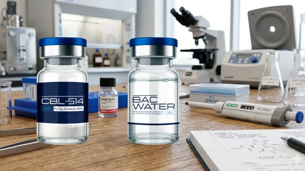
TECHNICAL AND SCIENTIFIC GUIDE CBL-514: Pharmacology, Clinical Evidence and Therapeutic Outlook Updated 29 July 2026 Intended audience: researchers,...
Read more...
1. Abstract Lyophilization, or freeze-drying, is the most effective method for stabilizing both synthetic and natural peptides. By...
Read more...
1. Abstract Ovágeno is a synthetic tripeptide (L-glutamic acid, L-aspartic acid, and L-proline) developed to act as a...
Read more...
1. Nature and Molecular Architecture AOD9604 is a synthetic peptide analog that mimics the C-terminal region of human...
Read more...
BIOPELABS delivers research-focused peptides with a clean, modern experience—clear product options, responsive support, and secure shopping. Explore our peptide catalog and find the right format for your needs today.
Disclaimer: The products offered by BiopeLabs are intended for research and laboratory use only. They are not approved for human consumption, therapeutic, diagnostic, or veterinary use. By purchasing from our website, you acknowledge that you are a qualified researcher or purchasing on behalf of a legitimate research institution and agree to use these products in accordance with all applicable laws and regulations.

1: 5-Amino-1MQ 10mg
2: 5-Amino-1MQ 50mg
3: ACE-031 1mg
4: ACTH 1-39 5mg
5: Adamax 5mg
6: Adamax 10mg
7: AHK-Cu 50mg
8: AICAR 50mg
9: AICAR 100mg
10: AOD-9604 5mg
11: AOD-9604 10mg
12: Alprostadil 20mcg
13: Amylin 5mg
14: Angiotensin (1–7) 5mg
15: ARA-290 10mg
16: Bimagrumab 5mg
17: Botulinum Toxin 100IU
18: BPC-157 10mg
19: BPC-157 20mg
20: BPC-157 5mg + TB-500 5mg
21: BPC-157 10mg + TB-500 10mg
22: Bronchogen 20mg
23: Cagrilintide 10mg
24: Cagrilintide 20mg
25: Cardiogen 20mg
26: Cartalax 20mg
27: CBL-514 10mg
28: CBL-514 20mg
29: CBL-514 30mg
30: Cerebrolysin 60mg
31: Cerlankin 2mg
32: CGRP (Calcitonin Gene-Related Peptide) 1mg
33: CJC-1295 with DAC 5mg
34: CJC-1295 with DAC 10mg
35: CJC-1295 with DAC 5mg + Ipamorelin 5mg
36: CJC-1295 without DAC 5mg
37: CJC-1295 without DAC 10mg
38: CJC-1295 without DAC 5mg + Ipamorelin 5mg
39: Chonluten 20mg
40: Cortagen 20mg
41: Crystagen 20mg
42: CT-1 Cardiotrophin-1 5mg
43: Dermorphin 5mg
44: Dihexa 5mg
45: DSIP 5mg
46: Dulaglutide 5mg
47: Elabela Apelin 5mg
48: Enfuvirtide 90mg
49: Epithalon 5mg
50: Epithalon 40mg
51: Epithalon 50mg
52: EPO 3000IU
53: FGF21 10mg
54: Follistatin-344 1mg
55: FOXO4 10mg
56: GHK-Cu 50mg
57: GHK-Cu 100mg
58: Ghrelin 10mg
59: GHRP-2 5mg
60: GHRP-6 5mg
61: Glucagon 1mg
62: Glutathione 1500mg
63: Gonadorelin Acetate 5mg
64: Goserelin 10mg
65: GLOW BPC-157 10mg + GHK-Cu 50mg + TB-500 10mg
66: HGH 191AA 15IU
67: HGH 191AA 36IU
68: HGH Fragment 176-191 10mg
69: HGH Fragment 176-191 15mg
70: HMG (Human Menopausal Gonadotropin) 75IU
71: Humanin 10mg
72: Hyaluronic Acid 5mg
73: Hexarelin Acetate 2mg
74: Hexarelin Acetate 5mg
75: IGF-1 LR3 1mg
76: Ipamorelin 5mg
77: Ipamorelin 10mg
78: Kisspeptin-10 5mg
79: Kisspeptin-10 10mg
80: KLOW BPC-157 10mg + GHK-Cu 50mg + TB-500 10mg + KPV 10mg
81: KPV 10mg
82: Leuprolide Acetate 10mg
83: Liraglutide 5mg
84: Livagen 20mg
85: LL-37 5mg
86: Lysyl Oxidase 30mg
87: Matrixyl 10mg
88: Mazdutide 10mg
89: Melanotan I 10mg
90: Melanotan II 10mg
91: Melatonin 10mg
92: MGF 2mg
93: MOTS-c 10mg
94: MOTS-c 40mg
95: NAD+ 100mg
96: NAD+ 500mg
97: NAD+ 1000mg
98: Nesiritide 5mg
99: Neuropeptide Y (NPY) 10mg
100: Ovagen 20mg
101: Oxytocin Acetate 5mg
102: Oxytocin Acetate 10mg
103: P21 5mg
104: P21 10mg
105: PACAP 5mg
106: Pancragen 20mg
107: PE 22-28 10mg
108: PEG-MGF 2mg
109: Pinealon 5mg
110: Pinealon 10mg
111: Pinealon 20mg
112: PNC-27 5mg
113: PNC-27 10mg
114: Prostamax 20mg
115: PT-141 10mg
116: PTH Fragments 10mg
117: Relamorelin 5mg
118: Retatrutide 10mg
119: Retatrutide 20mg
120: Retatrutide 30mg
121: Retatrutide 40mg
122: Retatrutide 60mg
123: Retatrutide 5mg + Cagrilintide 5mg
124: Selank 5mg
125: Selank 10mg
126: Semaglutide 5mg + Cagrilintide 5mg
127: Semax 5mg
128: Semax 10mg
129: Sermorelin 5mg
130: Sermorelin 10mg
131: SLU-PP-332 10mg
132: SNAP-8 10mg
133: SS-31 10mg
134: SS-31 50mg
135: Survodutide 10mg
136: TB-500 5mg
137: TB-500 10mg
138: TB-500 (FRAG) 10mg
139: Teduglutide 5mg
140: Teriparatide 10mg
141: Tesamorelin 5mg
142: Tesamorelin 10mg
143: Tesamorelin 20mg
144: Tesamorelin 5mg
145: Tesofensine 1mg
146: Testagen 20mg
147: Thymalin 10mg
148: Thymosin Alpha-1 5mg
149: Thymosin Alpha-1 10mg
150: Treprostinil 5mg
151: Vesugen 20mg
152: VIP 5mg
153: VIP 10mg
154: Vilon 20mg